Difference between revisions of "Main Page"
(→Update Log) |
(→Guides and Tutorials) |
||
| Line 40: | Line 40: | ||
# [[Surgery Protocol]] | # [[Surgery Protocol]] | ||
# [[Animal Behavior Guide]] | # [[Animal Behavior Guide]] | ||
| − | |||
| − | |||
| − | |||
| − | |||
== Update Log == | == Update Log == | ||
Revision as of 03:54, 18 January 2016
Welcome to Miniscope.org Wiki!
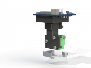
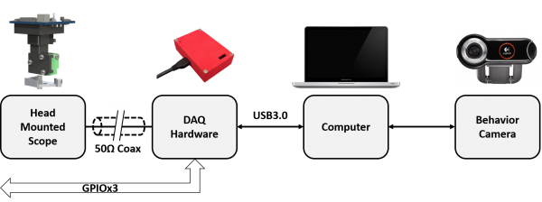
The miniature fluorescence microscope described here is based on a design pioneered by Mark Schnitzer's Lab at Standford and published in a paper in Nature Methods in 2011. It uses wide-field fluorescence imaging to record neural activity in awake, freely moving mice. The microscope introduced here (miniscope) has a mass of around 3 grams and uses only a single, flexible coaxial cable (0.3mm to 1.5mm diameter) to carry power, control signals, and imaging data. The goal of this wiki site is to provide a centralized location for sharing design files, source code, and other relevant information so that a community of users can share ideas and developments related to this important technology. The initial goal is to help disseminate this technology to the larger neuroscience community so that we can build a community of users that will continue to develop this technology and share on these developments. While the miniscope system described here is not an off-the-shelf commercial solution, we have focused on making it as easy as possible for a standard neuroscience lab to build and modify, requiring minimal soldering and hands on assembly. For more information please visit the Project Overview page. The Miniscope project and miniscope.org are still works in progress and will be routinely updated over the coming months and years. We hope you will contribute to this important process!
Contents
Current Status of Project
The Miniscope project is now in its third year of development at UCLA and has gone through two major revisions. The work and files available on this site are the most up-to-date public version of our system and will be updated frequently with improvements and new system features. Again, we hope that you will contribute to this development process! This wiki is designed for this very purpose.
Initial access to the miniscope.org wiki was enabled mid January, 2016.
Important: Using this system we have successfully imaged Hippocampal CA1, Subiculum, and Visual Cortex using 1.8mm and 2mm diameter GRIN lenses from Grintech. While thinner GRIN lenses should theoretically be compatible with our system we have limited our initial development to larger lenses due to supply and experimental constraints. We are now actively testing thinner lenses as well as pursuing multiple avenues of GRIN lens production (More information on GRIN lenses can be found here).
Links to information on miniscope subsystems
Discussion Board and FAQ
Guides and Tutorials
A key feature of this effort is to design miniscope systems that are easy to build and use. The guides below will walk you through component procurement, scope assembly, and software installation.
- Overview of System Components
- Part Procurement
- System Assembly
- Recommended Computer Specs
- Software and Firmware Setup
- Surgery Protocol
- Animal Behavior Guide
Update Log
- 01/14/2016
- Comments added to segmentation functions
- Added through-hole components for DAQ PCB on Master Parts List
- Added additional soldering tools on Master Parts List
- 01/13/2016
- Added basic surgery outline
- Added a picture guide for scope and Baseplate assembly
- 01/12/2016
- Finalizing of Miniscope Master Parts List
- 01/10/2016
- Upload of current version of all files and documents to Github
- 01/09/2016
- Added guide to programming firmware to DAQ PCB

