Difference between revisions of "Main Page"
From UCLA Miniscope
| Line 7: | Line 7: | ||
Our miniature fluorescence microscope uses wide-field fluorescence imaging to record neural activity and network structure in awake, freely behavin mice. The functional design of our microscope is similar to a table top wide-field fluorescence microscope with the following key differences: | Our miniature fluorescence microscope uses wide-field fluorescence imaging to record neural activity and network structure in awake, freely behavin mice. The functional design of our microscope is similar to a table top wide-field fluorescence microscope with the following key differences: | ||
| − | + | *All nonessential componets have been removed | |
| − | + | *The table top objective was replaced with an imaging objective GRadient Index of Refraction (GRIN) lens | |
| − | + | *The miniature fluorescence microscope does not use any infinity optics | |
| − | + | *The excitation light source is a super bright LED rather than a lamp or laser | |
| − | + | *The optical filters and dichroic mirror are custom cut to minimize their size and weight | |
| − | + | *A small CMOS imaging sensor is used to record emission light instead of a large CCD, emCCD, or sCMOS camera. | |
| − | + | *Adjustment of focal plane is done by moving imaging sensor not objective | |
Revision as of 13:33, 16 February 2015
UCLA MINIscope Resource
Welcome to the MINIscope Wiki page. If you do not have an account yet, please request one here.
Overview
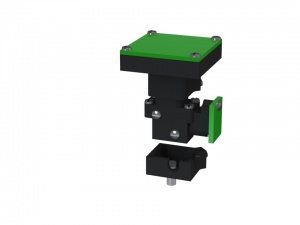
Our miniature fluorescence microscope uses wide-field fluorescence imaging to record neural activity and network structure in awake, freely behavin mice. The functional design of our microscope is similar to a table top wide-field fluorescence microscope with the following key differences:
- All nonessential componets have been removed
- The table top objective was replaced with an imaging objective GRadient Index of Refraction (GRIN) lens
- The miniature fluorescence microscope does not use any infinity optics
- The excitation light source is a super bright LED rather than a lamp or laser
- The optical filters and dichroic mirror are custom cut to minimize their size and weight
- A small CMOS imaging sensor is used to record emission light instead of a large CCD, emCCD, or sCMOS camera.
- Adjustment of focal plane is done by moving imaging sensor not objective
